Fibrocystic breast disease, also known as fibrocystic change or fibrocystic breasts, is a non-malignant condition commonly diagnosed in women. Your breasts may feel lumpy or rope-like, a condition that is more pronounced during the menstrual cycle. Although these changes can cause discomfort, it’s important to understand that fibrocystic breasts are not linked to a higher risk of breast cancer. The diagnosis of this condition is essential in differentiating between noncancerous lumps and potential malignancies.
Understanding the symptoms, which include breast pain, tenderness, and lumpiness, especially in the upper, outer area of your breasts, leads many women to consult a healthcare provider for diagnosis. In diagnosing fibrocystic breasts, healthcare professionals first perform a clinical breast exam. Following this, imaging tests such as mammography or ultrasound are often recommended to further evaluate the breast tissue. These diagnostic tools are valuable in providing a detailed view of the internal structure of your breasts, helping to distinguish between normal, fibrocystic tissue and any abnormalities that may require further investigation.
While mammograms are the standard screening tool for breast anomalies, ultrasound scans are also frequently utilized, particularly for younger women with dense breast tissue. This imaging technique complements mammograms by providing a clearer picture of cystic changes, which can be difficult to assess through mammography alone. If the diagnosis remains uncertain, additional procedures like fine-needle aspiration may be conducted to analyze breast cysts and alleviate discomfort. These diagnostic approaches are designed to provide reassurance and appropriate management of fibrocystic changes while ensuring that any potential concerns are addressed promptly and effectively.
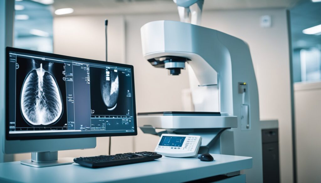
Diagnostic Imaging
In evaluating fibrocystic breast changes, imaging techniques like mammography and ultrasound examination are crucial. These methods help radiologists discern benign fibrocystic alterations from potentially malignant processes.
Mammography
When you undergo a mammogram, it involves a specialized form of X-ray to visualize the internal structure of your breast. Digital mammograms provide detailed images that can help discern dense breast tissue characteristic of fibrocystic changes from other concerns like breast cancer. It’s worth noting that mammography can sometimes be less effective if you have highly dense breasts, as the fibrous tissue and cysts can obscure clear imaging.
- Key Aspects of a Mammogram:
- Detects both calcifications and solid masses
- Can differentiate fibrocystic breast tissue from other abnormalities
- May include a clinical breast exam and breast self-exams for comprehensive assessment
Ultrasound Examination
During an ultrasound examination, sound waves are used to create images of your breast. This method is particularly good at determining if a lump is a fluid-filled breast cyst or a solid mass. It serves as a complementary tool to mammography, especially in cases of dense breast tissue where mammograms are less conclusive.
- Advantages of Ultrasound:
- Offers a clear image of breast cysts
- Helps in distinguishing between benign and potentially malignant lesions
- Non-invasive with no radiation exposure
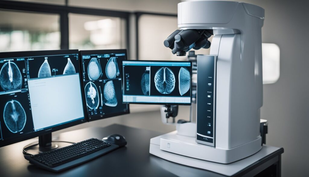
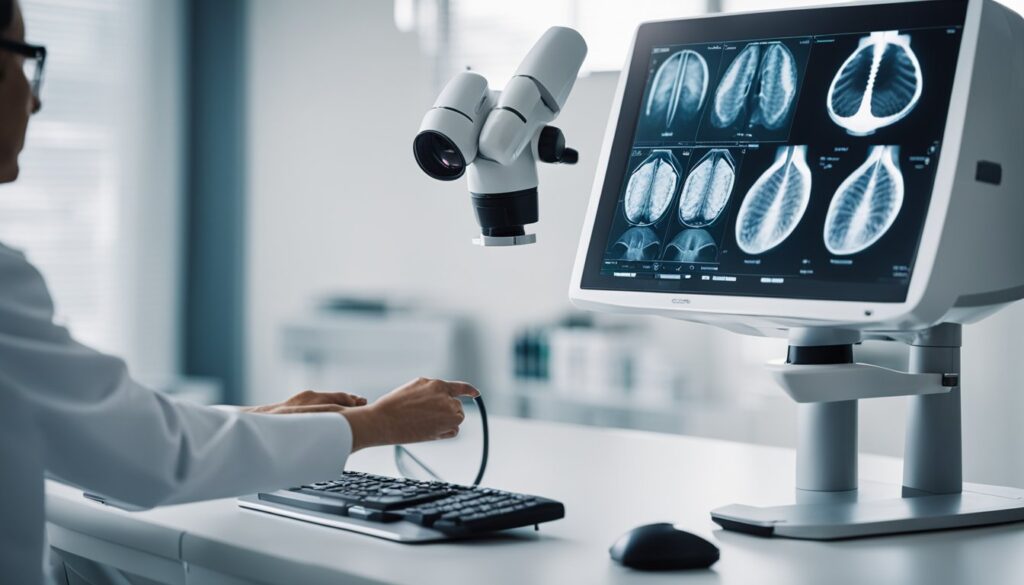
Comparative Imaging Techniques
Apart from mammography and ultrasound, additional imaging techniques like MRI (Magnetic Resonance Imaging) are sometimes utilized for a more comprehensive evaluation. Each imaging method has its strengths – for example, MRI can be more sensitive in some scenarios but is generally used as an adjunct to mammography, particularly in high-risk patients or those with very dense breasts.
- When to Consider Additional Imaging:
- If mammography or ultrasound results are inconclusive
- When there’s a personal or family history of breast cancer
- In the presence of symptoms despite negative mammogram and ultrasound
Your radiologist may use a combination of these imaging techniques to accurately assess and monitor fibrocystic breast changes.
Clinical Considerations and Management

When facing a diagnosis of fibrocystic breast disease, understanding the nuances of symptom evaluation, treatment options, and pathology is essential. It’s also important to consider lifestyle adjustments that may help with symptom management and risk reduction.
Symptom Evaluation
Your experience of fibrocystic breast disease can vary significantly with the menstrual cycle, often presenting with breast pain, swelling, and tenderness. These symptoms typically intensify during the premenopausal phase. Health professionals may consider hormonal changes and your detailed history to make a differential diagnosis, distinguishing fibrocystic changes from more serious conditions like carcinoma.
Treatment Approaches
Treatment for fibrocystic breast disease is often centered on symptom relief. Over-the-counter pain relievers such as acetaminophen or non-steroidal anti-inflammatory drugs may be recommended. In certain cases, hormone therapy including oral contraceptives or evening primrose oil, which is rich in fatty acids, can be beneficial. For persistent cysts, a fine-needle aspiration can provide both diagnosis and relief.
Pathology and Biopsies
In the presence of persistent or worrisome lumps, a breast biopsy might be necessary to rule out breast cancer. Types of biopsies include fine-needle aspiration and core needle biopsy. Pathological examinations look for signs of fibrosis, hyperplasia, and adenosis—conditions that fall under benign breast disease.
Lifestyle and Risk Reduction
You can engage in various lifestyle modifications to manage symptoms and potentially reduce risks. Reducing caffeine and salt intake, maintaining a diet rich in vitamins and low in fat, and regulating the consumption of alcohol are all suggested measures. For those experiencing discomfort, wearing a supportive or sports bra and using a heating pad might provide relief. The American Cancer Society recommends regular self-exams and screening as part of a proactive approach to breast health, especially in aging individuals.
Frequently Asked Questions
In diagnosing fibrocystic breast disease, identifying the symptoms and understanding the influence of diet and tissue changes are critical. Knowledge of the causes and duration of fibrocystic lumps can guide you through discerning it from breast cancer.
What are the common symptoms of fibrocystic breast disease?
You may notice lumpiness, tenderness, or pain in your breasts that can vary with your menstrual cycle when you have fibrocystic breast disease. Some breasts may feel thick and full, with areas of fibrosis that feel rubbery or firm.
How can diet influence fibrocystic breast disease?
Your diet can affect fibrocystic breast disease; reducing caffeine, fat, and carbohydrates might alleviate symptoms, while including a balanced intake of vitamins may help.
What does breast tissue thickening indicate in the context of fibrocystic breast conditions?
Thickening of breast tissue can often be a sign of fibrocystic changes. It’s your body’s response to hormones produced by your ovaries and can be distinguished from other conditions through detailed imaging like mammography or ultrasound.
What are the primary causes of fibrocystic breast disease?
Hormonal fluctuations, especially estrogen and progesterone, are primary causes of fibrocystic breast disease. These normal variations can lead to the development of fibrous tissue and cysts within the breast.
How long do fibrocystic lumps typically persist?
Fibrocystic lumps may persist for varying periods, some resolve after your menstrual cycle, while others can last longer. They are typically dynamic, changing in size and tenderness with your hormone cycles.
On what basis can fibrocystic breast disease be distinguished from breast cancer?
Fibrocystic breast disease and breast cancer can be distinguished based on imaging tests and possibly a biopsy. Fibrocystic changes are benign and usually present with definite patterns on mammograms and ultrasounds, while breast cancer may show irregular masses or calcifications.